Is magnification for you?
by Cherilyn G. Sheets, DDS
and Jacinthe M. Paquette, DDS
Technology is emerging in all fields and professions at a rapid pace. Dentistry is no exception. The changes are providing excitement and innovation for the dental professional and patient alike. Two challenges today are to be able to choose the advances that are most meaningful to your personal practice of dentistry and to progress rapidly through the necessary learning curves to make the technology useful on a daily basis. An important trend is the incorporation of higher levels of magnification and illumination into the operatory and the dental laboratory. This article will briefly review the history of this phenomenon and then look at the most popular current choices.
History
Most practicing dentists share a common experience. When they were dental students, the sight of "loupes" on the more senior faculty members indicated that the professor's eyesight needed assistance. In fact, a commonly expressed sentiment from dentists is that they feel they will not benefit from magnification because their eyes are still "good."
What has changed from "those days" is the realization that even the "best, normal vision" is not as good as vision assisted with magnification and illumination. The most significant indicator of this shift is that many dental schools immediately start their students with ocular magnification of 2.5x or greater. Preliminary studies show that even with 2.5x eyeglass magnification, dental students make 50 percent fewer errors in their tooth preparations. Increased visibility leads to in creased accuracy, re gard less of the clinician's age or physical attributes.
Current magnification options
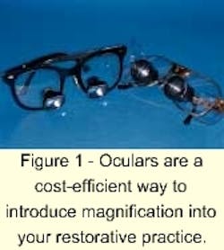
OCULARS - The most economical and rapid way to introduce magnification into your dental practice is the incorporation of magnification eyeglasses or oculars (Figure 1). Oculars can be manufactured with the eyeglass portion customized to the owner's prescriptive needs or with plain glass. The magnification lens can be at several magnification choices, ranging commonly from 2.5x to 6x.
Several companies cater to the needs of the dental profession and carry accessories such as headstraps, neck chains, plastic protective eye shields, flip-down light-curing shields, and frame design choice. Some practitioners have several pairs of oculars, allowing them to change magnification levels by changing glasses. A new, rapidly growing group of purchasers of ocular magnification sources is composed of dental hygienists and registered dental assistants.
The advantages of oculars include:
- a minimal to moderate cost for a pair of oculars
- a short learning curve
- mobility from operatory to operatory
- universal professional acceptance due to familiarity with the concept
The disadvantages are:
- a need for multiple oculars to change magnifications
- converging vision for the clinician's eyes, which is the same as with unaided vision and can be fatiguing during long appointments
- increasing weight of the oculars as magnifications increase
- lack of a light source to bring illumination to the magnified site
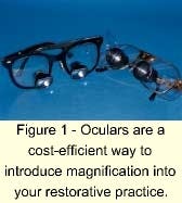
HEAD-MOUNTED ILLUMINATION SOURCE - An additional technological advance that often is combined with oculars is the illumination enhancement of a head-mounted light source (Figure 2). This provides increased lighting on the work surface, bringing the dynamic com bination of magnification and illumination to the oral cavity. The direction of the light is controlled by the direction of the operator's head. Many operators find that this additional component is well worth the investment.
The advantages of a head-mounted light source are:
- mobility from operatory to operatory
- increased visibility
- moderate cost
- the ability to purchase it independently from the magnification oculars
The disadvantages are:
- additional weight is added to the clinician's head as he/she leans forward and twists inward toward the mouth
- shadows are created by the singular light source
- the clinician is attached by a cord to an electrical outlet, limiting quick mobility to other sites in the office
- the head strap's impact upon the appearance of the clinician's hair, whether male or female, requires more frequent attention to personal grooming than is normally required in a workday
OCULAR/ILLUMINATION-ASSISTED VIDEO RECORDING FOR TEACHING - Variations on these two technologies are available. Some companies manufacture headsets that combine a headlamp with a video camera for teaching purposes. The goal is to bring illumination and video filming into the oral cavity to more closely create the operator's view.
Certainly, any advancement that can more effectively help students or postgraduate continuing-education participants see more realistic, live demonstrations is an advantage. These devices are an improvement over hand-held, remote-location cameras.
The advantages of ocular/illumination-assisted video recording are:
- a short to moderate learning curve
- moderate cost
- improved illumination and magnification for educational purposes
The disadvantages are:
- increased weight to the clinician's head
- jerky image movement due to changes in direction of the operator's head
- shadows from the unidirectional light source
- magnification for the operator limited to the projected image or from his/her personal oculars
INTRAORAL CAMERAS - Intraoral cameras certainly have brought magnification and illumination into the operatory for educational purposes with the patient and diagnostic purposes for the clinician. Unfortunately, the intraoral camera does not allow the clinician to have these benefits during the performance of dental procedures.
Technological advances allowing camera size to be condensed to fiberoptic proportions will allow more flexibility with bringing lifelike instructional video routinely into the oral cavity. The current systems still do not allow the clinician to actually have the advantages of magnification and illumination when working, even at the level experienced with oculars and head-mounted illumination systems.
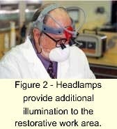
CLINICAL MICROSCOPES - Approximately 15 years ago, clinical microscopes were introduced into dentistry as an aid in the field of endodontics. The benefits of visibility due to "operator's view magnification" and working field illumination have revolutionized the specialty and discipline of endodontics. The average clinician, with the aid of technology, then was able to see defects and perform procedures at a competency level not generally achieved in prior decades.
Today, every endo dontic specialty program in the United States requires competency in the use of the clinical microscope prior to graduation. After the successes that were achieved in endodontics, periodontists began exploring the potential of microscopic-assisted periodontal plastic surgery. The pioneers in periodontics also spoke of the advantages of this technology for their specialty: improvement in accuracy, more delicate handling of tissues, less patient discomfort, im proved healing times, and more predictable esthetic and functional results.
It was only natural that eventually we would look at the possibility of using the clinical microscope for restorative dentistry. The results are basically the same - im provement in visibility from the magnification and illumination levels gives improved clinical results. General dentists and prosthodontists now can enjoy the same benefits experienced in other disciplines of dentistry if they incorporate the clinical microscope into their daily practice routine (Figure 3).
The advantages of the clinical microscope for the restorative dentist are:
- multiple choices of magnification with the twist of a knob, from 2x to 21x
- good posture, solving many traditional ergonomic problems associated with dental posture while chairside
- a more relaxed eye position, as the optical properties of viewing through a microscope eliminate the need to converge the eyes for focus
- illumination without shadows due to coaxial lighting from the microscope
- an apparent increase in fine motor skills due to increased visual acuity and visibility
- documentation benefits from attachments that allow 35mm or digital photography and video productions from the operator's view
The disadvantages of the clinical microscope are:
- a longer learning curve to master its use
- use of the microscope is predominately limited to one operatory
Cost is no longer a significant disadvantage to the clinical microscope, because some entry-level microscopes cost approximately the same amount as an intraoral camera. Ad dition ally, some companies provide leasing options for minimal monthly investments. As additional accessories are added to the microscope, the cost can increase. But these accessories are not necessary for some practitioners and can be added at a later time if desired.
Advantages of magnification and illumination

One question that is sometimes raised by dentists is: "How will acquiring tech nology to improve my visibility and ergo nomics im prove my bot tom line at the office?" (Figure 4). To that query, we have several potential answers, some from the patient's perspective and some from the dentist's perspective.
From the patient's perspective:
- When a dentist has invested in technology that helps provide a superior service, patients are pleased. When patients are pleased, they refer others to your practice who want the same type of services.
- Patients feel that dentistry must be precise. When a dentist uses additional sources of magnification and illumination, patients are assured that the dentist is committed to providing the most precise dentistry possible.
null
From the dentist's perspective:
- Magnification and illumination make it easier to do precise dentistry, which is professionally rewarding.
- Eye fatigue is reduced.
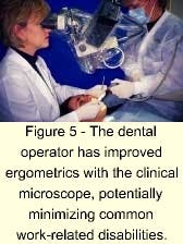
- Postural problems that can potentially lead to disability and perhaps a shortened professional life are minimized or eli minated when using the clinical microscope (Figure 5).
- Fewer costly remakes are required, because problems can be seen and eliminated at the preparation and impression stages of treatment.
- Overall patient treatment time may be minimized due to fewer problems with prosthesis fitting if magnification is used in the operatory and dental laboratory.
- An element of fun exists due to excitement at the quality of the work you and your staff can do, and the positive response that patients reflect back when they receivetreatment.
- Magnification and illumination help make it easier to provide quality care because you can see well. Providing quality care is profitable, because patients are willing to use discretionary dollars to purchase what they desire, and quality health care tops that list.
The next decade
There are times in our professional careers when the introduction of new technology changes our profession from that point forward. Dentists who have personally experienced the benefits of magnification and illumination have said their professional techniques are forever changed. It will be interesting to watch restorative dentistry over the next decade as magnification and illumination systems become commonplace in dental offices.