Part 2
The Magic Touch Concept presents a gentler way to conduct the oral exam.
Joseph J. Massad, DDS, and
William J. Davis, DDS, MS
Once we have determined the answers to the five questions posed in Part I (April 1999 Dental Economics), we can go further and begin our complete dental evaluation. We always ask the patient`s permission to do an oral examination (even though it is assumed that the patient is in the office for that reason). It becomes a much kinder and less invasive procedure when we ask the patient`s indulgence.
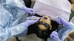
1) Notations With "The Magic Touch Concept," it is important to have a very light touch when retracting the lips to evaluate the oral cavity. While performing this examination, we systematically review each area of the oral cavity and read the findings aloud to the dental assistant as she makes notations. This is important because the patient can hear us review our findings and will, therefore, have a better sense of completeness.
Also, since the dental assistant will be putting these findings into the record, she will have a greater understanding of the patient`s condition. This will help the dental assistant to answer some of the patient`s concerns when the doctor is unavailable.
We are searching for any abnormal areas in the buccal mucosa, dorsum, lateral, and ventral surfaces of the tongue, soft and hard palate, gingiva (both attached and unattached), vestibular mucosa, and floor of the mouth. Any abnormalities noted should be listed and referred to at a later date in order to determine whether or not we have any pathology. For example, if we note an ulceration on the buccal area of the left cheek, we need to determine if it is simply due to a cheek bite or possibly other etiology. The ulceration may need to be biopsied in the future if the condition does not improve.
2) Tooth-by-tooth The next step is to review the teeth, tooth by tooth, beginning with tooth #1 and following through to tooth #32. (Although many practitioners like to start at the middle of the mouth, it is much better to begin at point A and proceed to point B. Thus, if we are distracted for any reason, it will be much easier to know where we were interrupted.) During this intraoral evaluation, we should have a full-mouth periapical radiographic series mounted on a view box and/or a panoramic X-ray. The radiographs verify our clinical findings during the tooth-by-tooth exam.
We find that tooth #1 is, for example, partially impacted, which is confirmed by the radiograph. Tooth #2 might disclose a mesial buccal cusp fracture with an existing occlusal mesial alloy. Tooth #3 may show an incomplete coronal fracture with an existing composite MOD restoration and visible recurrent decay. Tooth #4 could have a fractured buccal cusp with a short clinical crown. We continue this process until we have completed the tooth-by-tooth exam.
At this time, we discuss only these findings with the patient, avoiding any mention of treatment recommendations until we have compiled all appropriate information.
3) The gingiva Once we have completed the above review, we go back and evaluate the gingiva around each of the teeth. With a periodontal probe, we do a complete probing of both the buccal and lingual of each tooth in order to evaluate the depth of the pocket, any recession, and any attachment loss. Obviously, these findings are especially helpful before beginning any restorative procedure.
4) Occulsion We now observe the patient`s occlusion. We ask the patient to swallow in order to evaluate the centric occlusion. Many times, we will find that, when the patient closes, he may have a slide. We then try to relax the patient`s facial muscles by putting several cotton rolls between the maxillary and mandibular anterior teeth, asking him to slightly compress the cotton rolls for one minute. We now manipulate the patient`s mandible to obtain a repeatable motion in order to evaluate centric relation.
One way to manipulate the patient`s mandible is to secure the crown of the skull in the dentist`s diaphragm area and then perform bi-manual manipulation, as described by Dr. Peter Dawson. This will determine an unstrained, centric relation position. Often, we will find that only one or two teeth will make contact at this time. We may use articulating paper to visualize these observations and must make specific notations of them in the dental record.
In many patients the centric relation position is different than their centric occlusion. Knowing this, we will need to discuss the possibility of adjusting the tooth surfaces to develop a stable, repeatable position prior to any major restorative dentistry.
For the patient who is muscularly rigid and resistant to bi-manual manipulation, we generally use an acrylic anterior deprogrammer to facilitate a smooth, uninhibited movement. If this method is not successful initially, we instruct the patient to wear the deprogrammer during waking hours until his next appointment, at which time we repeat the procedure.
If a patient has any centric discrepancies, it is necessary to do a muscle palpation test to determine whether any symptoms are associated with this tooth-to-tooth prematurity. Our muscle palpation procedures should include palpating both anterior and posterior temporalis, the buccinators, the digastrics, both lateral and medial pterygoid muscles, and so forth. It is vital to thoroughly observe the temporomandibular joint at this time. Several standardized forms are available to guide the dentist through the procedure.
Patients who have severe worn dentition may either have occlusal problems or are possibly chronic bruxers - not discounting the possibility of a contributing medical condition. However, if this is not noted on the initial examination, we could have case failure after the treatment has been completed. Many patients may need to wear occlusal splint guards for most of their lives if they have this chronic situation.
5) Orthodontics Next, we evaluate the orthodontic condition of the teeth to determine the need for tooth repositioning. Orthognathic malalignment is a consideration that may necessitate surgery to enhance the final result. Many patients are resistant to surgical procedures. However, it is extremely vital that we review all aspects and explain them to the patient prior to discussing our treatment recommendations.
6) Endodontics The endodontic assessment is reviewed by palpating and percussing the teeth to determine whether or not there is any pulpal involvement. If we get any positive reaction to palpation or percussion, we use a pulp-testing device to make determinations about the vitality of the nerve. The cold test is still the standard test. Electric pulp stimulus follows for confirmation of nonresponsiveness to the cold test.
Any endodontic concerns will also be addressed prior to our planned treatment suggestion. All of these findings must be considered prior to discussing treatment options with a patient.
7) Models We then ask the patient`s permission to take a diagnostic models with a facebow and a centric relation record for proper mounting. At this same appointment, accurate maxillary and mandibular impressions are taken. We make sure that we maintain the proper vestibular height, vestibular borders of all teeth accurately. These impressions are poured up immediately to preserve the accuracy of the cast. A centric relation bite registration is taken by manipulating the patient`s jaw, and then an accurate facebow is completed.
These models then are mounted to complete the diagnostic workup. At this time, we correlate the full-mouth periapical radiographs and/or panoramic radiograph with the mounted diagnostic models and complete intraoral examination. The latter, of course, includes the periodontal charting, muscle palpation findings, and TMJ condition, as well as endodontic, periodontal, orthodontic, and orthognathic evaluations and the existing condition of the teeth and restorations. A systematic procedure simplifies and ensures a thorough examination.
8) Photographs It is extremely helpful to take intraoral photographs of the patient`s teeth, both in an open position and in tooth-to-tooth contact position. We also prefer to take both frontal and lateral views of the patient with his lips slightly touching. In order to do this, we have the patient swallow naturally and then come to a relaxed position prior to taking the photograph.
It is also helpful to photograph the patient`s entire facial features, including the eyes, nose, chin, and forehead. This gives us a perspective of the patient`s dentition around the facial mask. All of this becomes part of our permanent record and is what we use to assist us in providing treatment options for the patient.
At the time of the complete examination, only our initial findings are discussed with the patient so that he won`t feel too apprehensive in waiting for the consultation appointment. We do not go into details at this time, since we have not assimilated all the information. The patient is told that, once all the information has been reviewed, he should return to our office to discuss his treatment options.
We must make sure that the patient has given us sufficient information to assist us in providing a treatment option that is conducive to his particular needs. We can then recommend an ideal restorative procedure, taking into account the patient`s own specific concerns. It is our responsibility to convince a patient to want what he truly needs. Therefore, we will address several issues at the consultation appointment, including the original reason for his appointment, as well as the most ideal overall treatment recommendations for him. This may take care of some of the patient`s acute needs and then give us a chance to explain the long-term suggestions for total dental health throughout his lifetime.
This article is meant to be a brief overview. Each practice has to evaluate what its examination process includes.
Many patients will stop about halfway through treatment or may not want to complete all of the recommendations. However, it is our duty as dental physicians to ensure that a patient understands his specific treatment options and attempt to encourage him to follow through with total treatment in order to obtain ideal dental health.
We always let a patient know that nothing in life is perfect or permanent. However, we must strive for a healthy mouth and a clean, attractive smile. Most patients will relate to this and want to have all of the necessary procedures done. However, finances normally become an issue. In Part III of the exam, we will discuss how to reach a fair fee.
The dental forms used for graphics with this article were supplied by LeeMark Dental Products, (800) 800-3115.