Surfaces for operative restorations: A guideline for diagnosis and proper billing
Joseph P. Graskemper, DDS, JD, DABLM, FAGD
With the intrusion of third-party stakeholders, dental insurance companies, and government, there is a great need to have some basis for diagnosing the size of operative restorations upon which all can rely for fair and equitable patient care. In regard to billing, most dentists currently just rely on their individual feeling and ethical conscience as to what is fair when a restoration extends into another surface; there are no set guidelines to determine when to include another surface. We need a new paradigm that outlines a mutually fair and consistent manner in which to correctly diagnose and treat the number of surfaces involved in a restoration when a preparation extends into another unexpected surface of the tooth.
Prior to dental insurance, dentists were able to properly charge patients for the costs of a filling based on time, materials, and size. Now, for the most part, treatment is billed per an insurance fee schedule without consideration of the actual size of the restoration. For example, many Class II restorations may extend more facially or lingually past the marginal ridge area. Without established guidelines, dentists are left to arbitrarily decide whether to include the extended portion as another surface, and the insurance companies, of course, would rather pay only for two surfaces, when the restoration actually involves three. Insurance companies may even require verification that extra surfaces were actually involved during the restoration. The cost of verifying via an intraoral photograph is cost-prohibitive, however, since insurance companies most likely will not pay for the time, effort, and equipment/materials involved for the small amount of reimbursement.
I propose a guideline for dentists to be more united in the discussion of which surfaces are actually involved in the restoration of a tooth. The real question arises when a traditional Class II restorative preparation extends well into the facial or lingual area, thereby involving another surface. This can also be noted in large anterior restorations and large Class V restorations that wrap around to other surfaces. The insurance companies’ method of combining all surfaces to the least number of surfaces as a means of reducing payment is not proper or fair to the dentist, who has restored the tooth properly, or the patient, who wants fair reimbursement for the treatment received.
This new guideline must be fair to all professionals involved in the dental field: patients, dentists, and insurance companies. Every tooth is different, so caries removal may differ greatly depending on the skill and expertise of the dental provider.
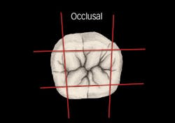
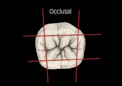
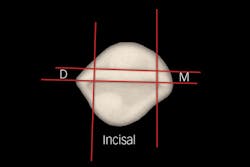
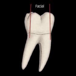
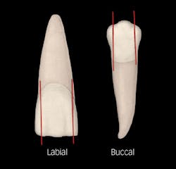
For proper diagnosis, treatment, and payment of services, each tooth is divided into four planes (two mesial-distally and two buccal-lingually), leaving nine surface areas as seen in Figure 1. Draw a line occusally and create a plane mesial-distally 1 mm lingually from the lingual edge of the mesial and distal contacts. Likewise, draw a second line/plane through the occlusal surface 1 mm buccally of the contacts. These planes should approximate the cusps of a molar 1 mm to 2 mm toward the center of the occlusal center or the width of the incisal edge of the anterior teeth. See Figure 2. Similarly, draw a line buccal-lingually 1 mm to 2 mm of the marginal ridge for a posterior tooth and the contact for an anterior tooth, sectioning the tooth into thirds. This should be approximately through the cusps of a molar 1 mm to 2 mm from the cusp tips from the marginal ridge. Or, you can divide an anterior tooth 1 mm to 2 mm from the mesial and distal incisal angles, cutting the tooth into thirds mesial-distally and creating nine sections of a tooth. The planes should extend through the labial-buccal and lingual surfaces as shown in Figures 3 and 4.
The illustrations show that as the tooth preparation extends into another plane, an added surface is encountered. As you go occlusally, mesially, or distally through the mesial or distal occlusal plane, another surface is encountered due to the undermining of the marginal ridges or the incisal corners for anterior teeth, which results in the need to restore another surface. Likewise, as you go buccally or lingually, you either undermine a cusp or are well beyond merely breaking the mesial or distal contact, and are actually into another surface. When preparing a large, deep facial abfraction/erosion, you would also encounter the mesial and/or distal plane, thus additional surfaces would require restoration.
This proposed guideline for dentists and insurers would foster a mutual understanding of the need to diagnose, treat, and receive proper remuneration for needed additional surfaces when the operative restoration extends beyond the traditional ideal preparation.
Joseph P. Graskemper, DDS, JD, DABLM, FAGD, is an associate clinical professor at Stony Brook School of Dental Medicine, where he teaches professionalism, ethics, and risk management. Dr. Graskemper has authored many peer-reviewed articles, lectured, published nationally and internationally, and recently published a book, Professional Responsibility in Dentistry: A Guide to Law and Ethics. He may be reached at [email protected] for comments or consultations.
