Ask Dr. Christensen
Gordon J. Christensen, DDS, MSD, PhD
Q One of my least desirable and successful clinical procedures is removable partial dentures. Many patients admit that they do not wear their partial dentures, except for esthetic reasons. It seems that I am always tightening clasps, and too often breaking clasps. Is there any way to make this undesirable treatment more acceptable for patients and for me?AYou are definitely not alone in expressing your dissatisfaction with removable partial denture (RPD) treatment. Most patients do not like their RPDs for the reasons you expressed. You can make RPDs much more acceptable for them by following some of these suggestions:Use of implants
One of the most important suggestions I can make concerning RPD fabrication is placing implants in strategic locations under the RPD. Placement of implants under removable partial dentures is well known to increase patient satisfaction by improving retention and stability. The implants are placed primarily for additional support and to provide some or all of the RPD retention. Although most dentists do not often accomplish this simple technique, I feel that implants should be used whenever patients can afford to have them placed. Where should the implants be placed?

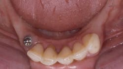
Fig. 1 — One implant has been placed in the almost-always present triangle of bone directly adjacent to remaining teeth.
Fig. 2 — Small-diameter implants can be placed in minimal remaining bone.
The implants should be placed as parallel as possible with the planned path of insertion and removal of the RPD. Depending on the attachment used, the parallelism necessary with the path of insertion varies. Spheres used as the abutments on the implants, with “O” ring rubber washers, allow the most variance from parallelism with the planned path of RPD insertion (Fig. 3).
Housings located to exactly coincide with the implant abutments are placed in the denture base resin of the RPD. There must be at least 4 mm of denture base material present from the tissue side of the denture to the artificial tooth to allow space for a housing to be placed.
The increase in patient satisfaction provided by implant placement under RPDs is due to the following improvements: clasps can be reduced in number or eliminated; and the retention provided by denture attachments such as ERAs and Locators or spheres and rubber washers is usually significantly more than that present with conventional clasps. When retention is reduced during service, it can be improved quickly and simply by placing a tighter plastic element in the attachment or replacing the rubber washer. Usually, implants can be placed in numerous sites depending on the locations of the remaining teeth.
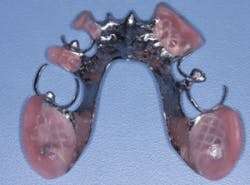
Fig. 3 — Rubber washers in housings, used as RPD retention, allow significant lack of parallelism of implants and still function well.
Keeping metal frameworks directly in contact with the gingival tissues
Many dental technicians place a spacer on the soft tissue represented on the working cast to allow space under the framework. This technique allows the framework to easily go into place without pressure on the soft tissue, but it also creates a food trap that collects food debris unless the partial is cleaned frequently. Also, air often escapes from under the denture, which is disagreeable to the patient. I suggest the framework should rest directly on the soft tissue, and that there should be NO relief of the stone cast (Fig. 4). When using this concept, a sore spot will occasionally occur after seating the RPD. The sore spot can be eliminated quickly and easily with various pressure evaluation materials, such as Fit Checker from GC, to help find the spot and eliminate the pressure. This is a small concession for the elimination of food entrapment and air escape.
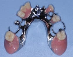
Fig. 4 — The stone cast has not been relieved for this RPD, and the metal framework rests directly on the soft tissue. This technique reduces metal bulk and reduces food impaction around the RPD.
Using thin frameworks
Typical RPD frameworks are often made thick and bulky, apparently in an attempt to reduce breakage. The resulting bulky and large RPD is objectionable to the patient. The clasps are rigid and cannot flex. When the clasps of such frameworks are tightened, the partial frequently will not fit back easily into the mouth. I suggest that RPD frameworks should be made thin to allow flexibility (Fig. 5). A thin RPD framework can be slightly flexed when held in your hands and force is applied. Of course, the negative aspect of thin frameworks is breakage, but this is so infrequent that it should not be a major deterrent. Patients appreciate thin and flexible frameworks because the RPD does not have the peculiar, objectionable, large, foreign object feeling of a thick framework.
Making clasps thin and flexible
Thick, nonflexible clasps are unsightly, cannot be easily tightened, and are bulky and uncomfortable for patients, thus contributing to patients’ reluctance to wear an RPD. I suggest making thin, flexible clasps that easily spring into undercuts (Fig. 7). Most laboratory technicians need instruction to make thin, flexible clasps, since they usually make them thick in an attempt to avoid breakage. In my experience, thin, flexible clasps do not require significant tightening, they retain RPDs well, and they satisfy patients far more often.
Fig. 5 — This metal framework has purposely been made thin and flexible, reducing rigidity and bulk and improving patient acceptance.
Smooth RPD frameworks
Some labs make the external surfaces of frameworks rough, apparently in an attempt to make them similar to the irregularities on natural gingival tissue. In my experience, patients prefer smooth external RPD surfaces that can be polished to a high shine with extremely smooth surfaces (Fig. 6).
Fig. 6 — Note the smooth external surface on this RPD framework. Most patients prefer this type of surface to the rough surface often placed on RPDs by technicians.
Lingual clasps
Historically, most retentive and supportive clasps for RPDs have wrapped around teeth, and they show metal on the facial surfaces of the teeth. Some clasps, such as “I” bars, locate a vertical bar of metal on the facial surfaces of the supportive teeth. Placing clasps in any area where the patient can see them negatively influences patient acceptance. Additionally, observers can easily see the clasps, which immediately says that the patient is wearing an RPD. Reducing or eliminating this metal display can be accomplished simply by using the following procedure:
- Identify the locations for the RPD design where retention is necessary.
- Prepare occlusal rests in the teeth involved where retention is necessary. The rests should be at least 1 mm deep and should eliminate the possibility that the framework can move out of the rest seat toward the facial or lingual.
- If the rests must extend into dentin, a resin-based composite restoration should be placed in the deepest portion of the rest, and the patient should place fluoride gel onto the rest seat metal at least once a day. (An example is Colgate PreviDent 5000 Plus fluoride gel.) Potential retentive undercut areas should be identified on the lingual tooth surfaces of the affected teeth.
- If gingival tissue blocks the entry of a clasp into the undercut, a small piece of soft-tissue should be removed with an electrosurgery unit or a laser, allowing the tip of the clasp to enter the undercut.
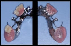
Fig. 7 — Note the thin and flexible clasps on this RPD. Such clasps allow easy insertion and removal of the RPD, spring easily in and out of undercuts without distortion, and are less objectionable esthetically for patients.
The framework is designed with minimal or no facial clasp display (Fig. 7).
Flexible denture base materials for some RPDs
This type of RPD is not the restoration of choice for most patients, because the RPD may sink into soft tissue, break, or cause occlusion challenges by collapsing the occlusion and causing opposing tooth extrusion. Although condemned by some dentists, in my opinion, flexible, non-metal-containing RPDs serve some purposes well.
Their use is acceptable for treated periodontal cases with somewhat mobile teeth, for those patients not suitable for implants, or for those patients who have difficulty wearing conventional metal framework containing RPDs. In my opinion, they are a satisfactory clinical solution if they’re accompanied with adequate informed consent and patient education.
In summary, current excellent materials, placement of implants, and improved patient-friendly techniques can make RPDs acceptable treatment. I suggest that you look into the various suggestions I have made and implement those that fit your patient population.
Our newest DVD, “Predictable Removable Partial Dentures” (Item# V2551), illustrates many of the concepts we have identified in this article, and shows me, Dr. Christensen, in live, close-up video accomplishing the procedures. Visit our website www.pccdental.com or call Practical Clinical Courses at 800-223-6569 for additional details.
Dr. Christensen is a practicing prosthodontist in Provo, Utah. He is the founder and director of Practical Clinical Courses, an international continuing-education organization initiated in 1981 for dental professionals. Dr. Christensen is a cofounder (with his wife, Rella) and senior consultant of CLINICIANS REPORT (formerly Clinical Research Associates).
Past DE Issues